The aim of this work is to underline the effectiveness of a protocol which involves the use of Platelet-rich plasma as a
grafting material in the implant rehabilitation.
The Authors included in the study protocol a cohort of 27 patients requiring
maxillary sinus lift, before implant-prosthetic rehabilitation, by using PRP in
combination with autogenous bone, anorganic bone material and organic bone
substitutes.
The treated patients were included in a follow-up plan, which established
clinical and radiological examinations on the day after surgery and six months
later.
The success of the implant-prosthetic rehabilitation, defined as primary
stability and radiographic integration at the moment of the implant placement,
was of 100% (all 27 patients out of 27 included in the described protocol).
KEYWORDS
PRP, SINUS LIFT, TISSUE REGENERATION, GROWTH
FACTORS.
Fig. 1 Dentascan of one of the most representative cases: a 49-year-old female patient. It is possible to observe a great loss of alveolar bone in teeth 16 and 17. |
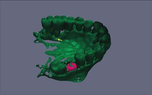 Fig. 2 A detail showing the three-dimensional reconstruction of the patient?s maxillary arch. The yellow arrow indicates oroantral communication. |
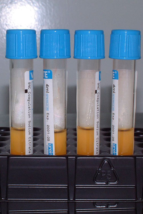 Fig. 3 4 tubes containing 4 ml of PRP each. |
 Fig. 4 Placement of two implants in teeth 16 and 17, subject to endosinusal injection of liquid PRP. The implant placement was followed by PRP packing, activated with Calcium Chloride + Bio-Oss. |
 Fig. 5 A detail showing the Orthopantomogram performed the day after surgery. |
Fig. 6 Tomogram showing x-ray integration and the increased density of the peri-implant bone, six months after surgery. |
Fig. 7 Three-dimensional view that allows to examine the newly-formed bone in the peri-implant area. |
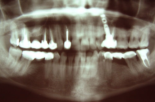 Fig. 8 Orthopantomogram performed before surgery in a 45-year-old female patient included in the protocol. |
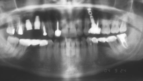 Fig. 9 Post-surgery control Orthopantomogram in one of the patients included in the protocol. Two implants were placed in teeth 15 and 16, subject to PRP packing in the sinusal region. |
 Fig. 10 Orthopantomogram performed six months after surgery. It is possible to observe the presence of newly-formed bone in the peri-implant area. |
 Fig. 11 Tomograms of the dentascan performed six months after surgery. |
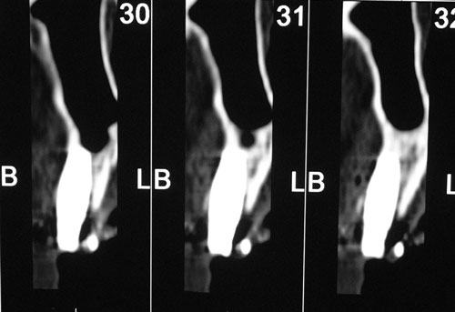 Fig. 12 Tomograms of the dentascan performed six months after surgery. |
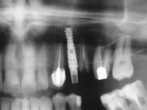 Fig. 13 A detail of the pre-surgery Orthopantomogram in a 58-year-old male patient, who was included in our protocol. |
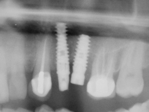 Fig. 14 A detail of the Orthopantomogram performed three months after implant placement in tooth 25. |
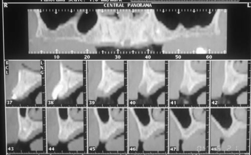 Fig. 15 DentaScan performed before surgery in a 75-year-old patient. |
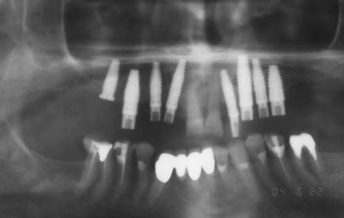 Fig. 16 Post-surgery Orthopantomogram performed after placement of eight implants, seven with immediate load and one with delayed load (tooth 15). |
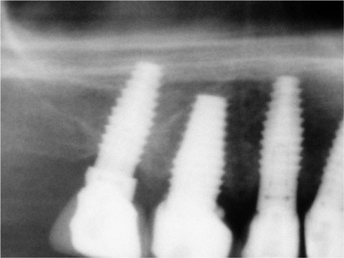 Fig. 17 A detail of the control Orthopantomogram performed three years after surgery. |
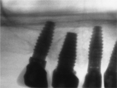 Fig. 18 Negative of the previous picture. |